News from Authors: Dear students and teachers, Now the blog is full on and all are welcome to present their article in this blog related to neurobiology. please provide your valuable feedback on this. With regards Biosaints

Bax, Par-6 and aPKC is not required for axon and dendrite specification

Overexpression of Baz or Par-6, aPKC does not have any effect on axon morphology or number in Drosophila melanogaster (fruit fly). New study shown by Melissa M Rolls from penn state and Chris Q Doe from univ of oregon.

GSK3b- The master regulator of neural progenitor homeostasis

GSK-3 deletion have resulted in suppression of both intermediate neural progenitors and postmitotic neurons generation and massive hyperproliferation of neural progenitors along the entire neuraxis.

EphrinBs reverse signalling and control dendrite morphologies

EphrinB signalling pathway regulates the dendrite morphology and number of spines.

No need of Protein synthesis in distal axon for growth cone response to the cues

New study have shown that protein synthesis is not required for growth cone response to guidance cues. The study was investigated over chick neuron by researchers from univ of Minneapolis, Minneapolis.

SOX 2 gene decides the neural stem cells fate

New study shown that SOX2 maintains the potential for neural crest stem cells to become neurons in the PNS. The research was led by Dr.Alexey Terskikh at Sanford-Burnham Medical Research Institute (Sanford-Burnham)

Oct 30, 2011

Negative regulators of Neuronal polarity

Negative regulators of neuronal polarity means the molecules will inhibit other downstream or upstream molecules which are involved in controlling the Cytoskeleton formation, stability, endocytosis etc.

PTEN (Phosphatase and tensin homolog)

It is a lipid phosphatase protein which counteracts the enzyme PI3 K (PI3 kinase) by converting PIP3 formed by PI3K into PIP2. Overexpression of this molecule will lead to prevention of PAR 3 localization to neurites and inhibits axon formation. If the PTEN expression is downregulated using SiRNA, then all the neurites becomes axons and there will reduction in dendrite formation. From this we can conclude that PTEN is important for the growth specific single axon. PAR 3 (Drosophila epithelia and neuroblasts) actually directly interacts with PTEN and this property leads to negative feedback loop. Where the PI3Kinase will recruit PAR 3 which in turn brings the PTEN molecule, thus leading to maintenance of PIP3 concentration in axon or PIP2 formed will bind to actin associated proteins such as WASP and regulate the actin formation.

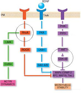
PTEN is associated with PI3K and GSK3β; GSK3β inhibits PTEN by Phosphorylating at its C terminus. Until GSK3β is active, the PTEN activity is downregulated and therefore upregulation of PTEN happens by GSK3β inhibition which might provide a negative feedback regulation by again counteracting at PI3K activity.

Rho A (Rho GTPase Family)

Rho A is a Rho GTPase family protein which has the negative regulation role in the axon stability. Rho A negatively regulates the neurite outgrowth and modulating its signaling pathway will lead to axon regeneration in the central nervous system during injury.  ROCK which is a Rho kinase effector accompanies Rho A in regulating axon growth and actin dynamics during neuritogenesis of cultured cerebellar granule neurons. Rho A-ROCK pathway with profilin II regulates actin and microtubule stability. Still Rho A knock mice is not created inorder to analysis the function of Rho A in neuronal polarity.

GSK3β (Glycogen synthase kinase 3β)

GSK3β is negative regulator of CRMP -2 (collapsin regulated mediator protein-2) which is GSK3β substrate responsible for microtubule polymerization. GSK3β is regulated by Akt protein which is activated by the PIP3 produced by PI3 Kinase. The protein not involved in establishment of neuronal polarity, but also plays a vital role in maintenance of neuronal polarity. CRMP-2 will be phosphorylated at thr-514 by GSK3β thereby making it unable to bind with tubulin dimer and NUMB. Overexpression of active GSK3β will lead to prevention of axon formation, whereas in inactive form it will lead to multiple axon formation. It also inactivates the MAP (microtubule associated proteins) protein by Phosphorylating MAP1B and APC (adenomatous polyposis coil).

Oct 13, 2011

Neuronal Polarity Introduction

The formation of single axon and multiple dendrites in a neural cell through distribution of specific functions to discrete cellular domains.

The neurons undergo complex morphological rearrangements to assemble into neuronal circuits and propagate signals. Hence they start as a round neuronal spheres and gradually adopting a complex morphology by forming one long axon and several shorted dendrites to eventually connect to target cell or other neurons via synapses.  The central importance for the axon formation and the neuronal polarity is the specialized, highly motile cellular compartment at the tips of the growing axons called growth cone. 

Different stages of neuronal polarity 


Stage 1: Neuronal development starts with round spheres that spread a lamellipodium around the cell body


Stage 2: Then transform into cells containing several neuritis, which are decorated with dynamic growth cones at their tips.

Stage 3: Neurites at this early developmental stage show characteristic alternations of growth and retraction. The major polarity event is when one of these equally long neurites starts to grow rapidly to become the axon.

Stage 4: The next step is the morphological development of the remaining short neurites into dendrites.

Stage 5-6: Here Functional polarization of axons and dendrites, including synapse formation happens and dendritic spines are formed at later stages.

Cytoskeleton Role in Neuronal polarity

Cytoskeleton plays a vital role in both establishing as well as maintaining polarity in neurons. Basically actin filaments and microtubules have the functional properties which make them uniquely suited to determine and regulate polarity in neurons as well as in other polarizing cells.

Two basic characteristic features of Actin filament and microtubules in polarity:

  1.        The Microtubules rapidly convert the molecular signals into structural changes modulating cell shape.
  2.           They possess an intrinsic polarity.

Growth cones support the growth by sensing environmental cues and transducing those signals to the cytoskeletons.

The structure (axonal growth cone) is basically composed of a central region filled with organelles and microtubules, whereas the pheripheral region is highly dynamic, actin rich area containing lamellipodia and filopodia.


it’s shown here that the barbed ends of the actin filament which is oriented towards the rim and latter pointed ends are towards the base of the growth cone. We can find that G-actin subunits are continuously incorporated into the barbed end while the other pointed end is found to be dissociated which is basically resulting in a “treadmilling” of F actin and regulation of growth cone dynamics.


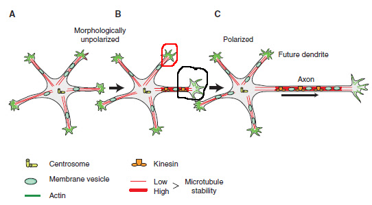
In the initial establishment of the polarity, rearrangement of the actin cytoskeleton and microtubules is very important. There is an enhanced growth cone dynamics shown by the future axon before the occurrence of morphological polarization (circled in black).  Even the future dendrites are not growing properly at that stage, therefore it have a static growth cone with a rigid actin cytoskeleton (circled in red). If we pharmacologically depolymerase the actin cytoskeleton, then the non growing dendrites will be transformed into growing axons. It suggests that the actin filaments of the future dendritic growth cones form a barrier for the protrusion of microtubules, whereas the axonal growth cone contains an actin structure permissive for the microtubule protrusion.


Microtubules also actively participate in the neuronal polarization and they are more stable in the axonal region compared to the minor neurites. The stabilization of microtubules is far more sufficient to induce axon formation.      

Oct 12, 2011

Actin and Microtubules regulators


Actin regulating proteins
Basically the actin dynamics are regulated by actin nucleating, severing, branching and bundling proteins.

Actin nucleating proteins are Arp 2/3 complex (Lamellipodia) and formins (Filopodia). The Arp 2/3 complex is also very important in the formation of Filopodia as well as dispensable for actin organization in neuronal growth cones. 
Growth cone dynamics regulators (directly modulate actin dynamics):
  • WAVE (wiskott Aldrich syndrome protein [WASP] family verprolin homologous protein)
  • Ena/VASP (Vasodilator stimulated phosphoprotein)
  • Profilin
  • ADF (actin depolymerization factor)/ cofilin
WAVE protein is localized in the Lamellipodia and is part of the WAVE complex of the proteins that act together to regulate actin polymerization in Lamellipodia. Basically the WAVE pathway regulates the actin polymerization through the Arp 2/3 or Profilin and promotes axon growth. 

A component of WAVE complex called Nap 1 (Nck associated protein 1) which is important in cortical neuronal differentiation as well as proper extension of axon.

The Ena/VASP proteins are localized at the tips of the Lamellipodia and Filopodia, which will accelerate the actin polymerization by their anticapping activity and bundle actin filaments. Absence of these proteins will lead to aberrant actin bundling and failure of Filopodia formation. Therefore formation of neurites in the absences of these proteins is not possible, hence it’s considered important in the neuron formation.
The role of Ena/ VASP signaling in axon formation appears to be conserved throughout the species. Additionally, Ena/VASP proteins recruit the actin regulator profilin, but the physiological relevance of this interaction is still not clear.

Profilin localizes to the leading edges of the growth cones and enhances the formatting of ATP bound G actin monomers that are incorporated into actin barbed ends. Ablation of isoform Profilin IIa protein leads to destabilization of actin cytoskeleton as well as increased in the number and length of the neurites.

But it was investigated in the Profilin Knockout mice, that polarization has taken place normally. It shows that Profilin I is compensating the function of Profilin II, But Profilin I knockout mice are embryonical lethal. 

Cofilin is an actin severing proteins which are also implicated in the neuronal polarity. ADF and cofilin І which are expressed in brain is found to be abundant in the neuronal growth cones. They bind mostly to the ADP-actin rather than ATP actin which results in their association with the pointed end of actin filaments and promoting actin depolymerization.  Both the proteins are regulated by Phosphorylation and phosphorylated form is mostly abundant in the cellular forms. The cofilin proteins are active during axonal growth cones compared to non growing future dendritic growth cones.


Microtubule regulating proteins

CLIP (Cytoplasmic linker proteins) and CLASPs (CLIP associated proteins) regulated the microtubule dynamics.  They act as MT growth promoting and also MT stabilizing protein.

CRMP 2 (collapsin response mediator protein 2) binds to the free tubulin and promotes their capacity to bind to the microtubules. CRMP2 is inhibited by GSK 3β Phosphorylation and PTEN. WAVE complex helps the transportation CRMP2 on kinesin molecule.

Stathmin/Op18 is the MT destabilizing proteins, which can be inactivated inorder to attain MT stability and is necessary for the specification of the nascent axon.  

Neuronal polarity regulation


The hippocampal neurons form a spikes and small proturusion veils at stage 1. This is stage where the neuron stem cell breaks out from the cell cycle.


Then the truncated protrusions develop into a several neurites at stage 2.  
At stage 3, All the neurites are roughly equal in length, in which only one neurite will start to break the initial morphology symmetry and grows at rapid rate and thus they eshtablish polarity.

The extracellular cues can guide which neurite to grow as axon. The extracellular cues are highly concentrated over the stage 2 immature neurites. Well the guidance happens through the help of downstream signalling molecules present within the neurites. PI 3 kinase and its lipid products such as PIP3 determines and maintains the internal polarity.  PI 3 kinase activity is localized at the tip of the newly specificed growing axon and its activated when its get the signal from the extracellular cues.

The Signalling cascades helps the centrosome to position at one of the neurite and these centrosome position is responsible for the growth of a axon, because it guides the microtubules to move in one particular neurite. If a neuron is having multiple centrosomes, then it will lead to formation of multiple axons. From this its well understood that centrosome is indirectly responsible for the axon formation, since its controlled by PI 3 kinase, cdc 42 and other regulatory molecules.
Soon after the axon starts it rapid growth action, the other neurites enlongates and acquire the charactersitics of dendrites which is at stage 4.

Finally the neurons form the synaptic contacts and eshtablish a neuronal network at stage 5.
So at stage 2, There happens to be two kinds of feedback signals which is very balanced. They are positive and negative feedback signals. In this negative regulations, the actin dynamics decrease and microtubule catastrophe occurs, whereas in positive regulation, actin dynamics increases and microtubule assembly occurs. Hence the neurites are found to be of same length and it wont grow unless some extracellular cues triggers the downstream regulating molecules. When balance is broken, then the axon growth occurs, where the postive feedback plays a major role than the negative feedback loop.

PIP3, Akt and GSK 3β signaling cascade determines the fate of the axon growth. When the receptor gets activated or triggered by some extracellular cues like netrins or BDNF, then it’s the Ras, which is a G protein activates the PI-3 kinase. The PI-3 kinase converts the lipid membrane molecule PIP2 to PIP3 which activates the PDK 1 (Phosphoinositide dependent kinase 1). The PDK 1 and other kinases such as integrin linked kinase phosphorylates the kinase Akt protein (PKB) at threonine 308 and serine 473. The phosphorylated Akt protein will inactivate the GSK 3β by Phosphorylation.

GSK 3β will activate the regulator proteins which is will be responsible for the microtubule catastrophe. Hence the activated GSK 3β or overexpression of GSK 3β will not allow the formation of axon, whereas knockout of GSK 3β gene have lead to formation of multiple axon. Therefore, the GSK 3β will be found inactive at the tips of the growing axon or growth cone.  

RAP 1B (ras related protein 1B), which is ras superfamily GTPase localize at the tips of the axon before the cdc 42 and PAR complex accumulates. It is said that the accumulation RAP 1 protein in one particular neurite is responsible for the axon specification and recruitment of other proteins for axonal growth.  The TrkA A receptor is activated by the extracellular cues, which will consequently recruits the GEFs of RAP 1 and adaptor protein (CRK) and then stimulates the RAP1.

Rho GTPase: Rac 1 and cdc 42 activation will lead to the elongation of the neurite, whereas in the case of Rho A activation will lead to the inhibition of the neurite outgrowth. Certain Rho GTPase effectors also influence in the neurite outgrowth such as P35, PAK, MRCK, N- WASP and IQGAP3. There are regulators of Rac 1: TIAM 1 and STEF (SIF and TIAM 1 like exchange factors), which regulates the upstream molecules of it. 

PAR complex: PAR 3 and PAR 6 with aPKC will form a complex with cdc42. The PAR complex will activate the Rac 1 protein through the TIAM 1 and STEF. 


This picture shows how the PAR complex gets localized. They are transported from the cell body to the growing axon by the motor protein kinesin 2.  


Oct 11, 2011

PAR protein in axon specification and neuronal polarity


Par (for partition defective) proteins are evolutionary conserved proteins, which are involved in regulating axon specification and establishment of neuronal polarity.  These proteins are first discovered in the C.elegans, which has 6 par genes named as par1-6.

s. no
Par proteins
In C.elegans
Drosophila homolog
Mammalian homolog
Involvement in neuronal polarity?
1
Par1
Serine/Threonine kinase
Par 1
MARK kinase
SAD kinase
Yes
2
Par2
Zinc finger and ATP binding motif (ubiquitination pathway)
Not conserved
Not conserved
unknown
3
Par3
Scaffold (PDZ domains)
Bazooka
mPar3 (also called as ASIP PHIP)
Yes
4
Par4
Serine/Threonine kinase
Par4
LKB1
Yes
5
Par5
14-3-3 protein
Par5
14-3-3 protein
Unknown
6
Par6
Scaffold
Par6
mPar6  
Yes

Par3/Par6/aPKC complex in neuronal polarity

Par3/Par6/aPKC complex plays a crucial role in axon specification and polarity establishment in a cultured hippocampal neuron. These complexes (Par3/Par6) are selectively enriched at tip of the future axon, inorder to make the cell polarized. If the complex expression is inhibited, then the neurite differentiating to axon and dendrite does not take place. If the complex is over expressed, then multiple axon or dendrite is formed.

The par3 and par6 are scaffold proteins, where Par 3 interacts with Par6 and with Tiam1 (T-lymphoma invasion and metastasis 1) and GNEF (Guanine nucleotide exchange factor) for Rac. Par6 forms a stable complex with aPKC (protein kinase C) and contains a semi- cdc42/Rac interactive binding (CRIB) domain that specifically binds to the active GTP bound form of the small GTPases, Cdc42 and Rac1 (central regulators of actin cytoskeletal dynamics).    

It also regulation of these par complexes is responsible for the control in morphogenesis of dendritic spines. 

Cdc42 which is activated by the PIP3 will initiate the positive feedback loop by binding to the Par6 protein.  

The par3/par6/aPKC complex will then activate Rac GEF which has STEF/ Tiam1 protein bind to it. This activated Rac complex will activate the Rac, a small GTPase protein and finally triggers the PI3 kinase. PI3 kinase is the important enzyme in neuronal polarity and axonal specification because it converts the PIP2 to PIP3, a molecule responsible for activation of other proteins playing a vital role in the axon growth. 

The par 3/ par6 complex will also inhibit the GSK3β which is responsible for the inhibition of CRMP2, Tubulin, APC and microtubule associated proteins (MAPs). 


Definitions


Axon Specification

In establishing a polarized neuron, the initial event is the specification of a single axon where one of the neurite will grow into a axon.